Review sheet 30 anatomy of the heart – Prepare to embark on an anatomical journey as we delve into the intricacies of the heart. Review Sheet 30 serves as your guide, providing an in-depth examination of this vital organ, its structures, and its pivotal role in our circulatory system.
From the heart’s external features to its intricate internal components, we will explore the pathways of blood flow and the electrical conduction system that governs its rhythmic beating. This comprehensive overview promises to deepen your understanding of cardiovascular anatomy, equipping you with a solid foundation for further exploration.
Heart Anatomy Overview: Review Sheet 30 Anatomy Of The Heart

The heart is a vital organ responsible for pumping blood throughout the body, delivering oxygen and nutrients to tissues and removing waste products. Located in the thoracic cavity, slightly to the left of the midline, the heart is a muscular structure enclosed within a protective sac called the pericardium.
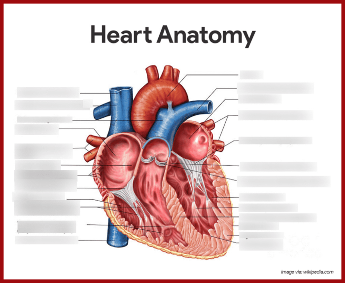
Internally, the heart consists of four chambers: two atria (upper chambers) and two ventricles (lower chambers). The right atrium receives deoxygenated blood from the body and pumps it to the right ventricle. The right ventricle then pumps the blood to the lungs for oxygenation.
Oxygenated blood returns to the heart via the left atrium and is pumped by the left ventricle to the rest of the body.
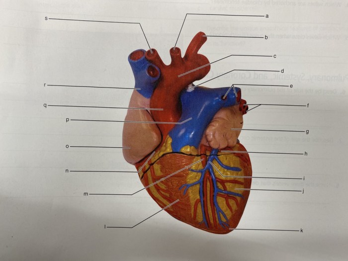
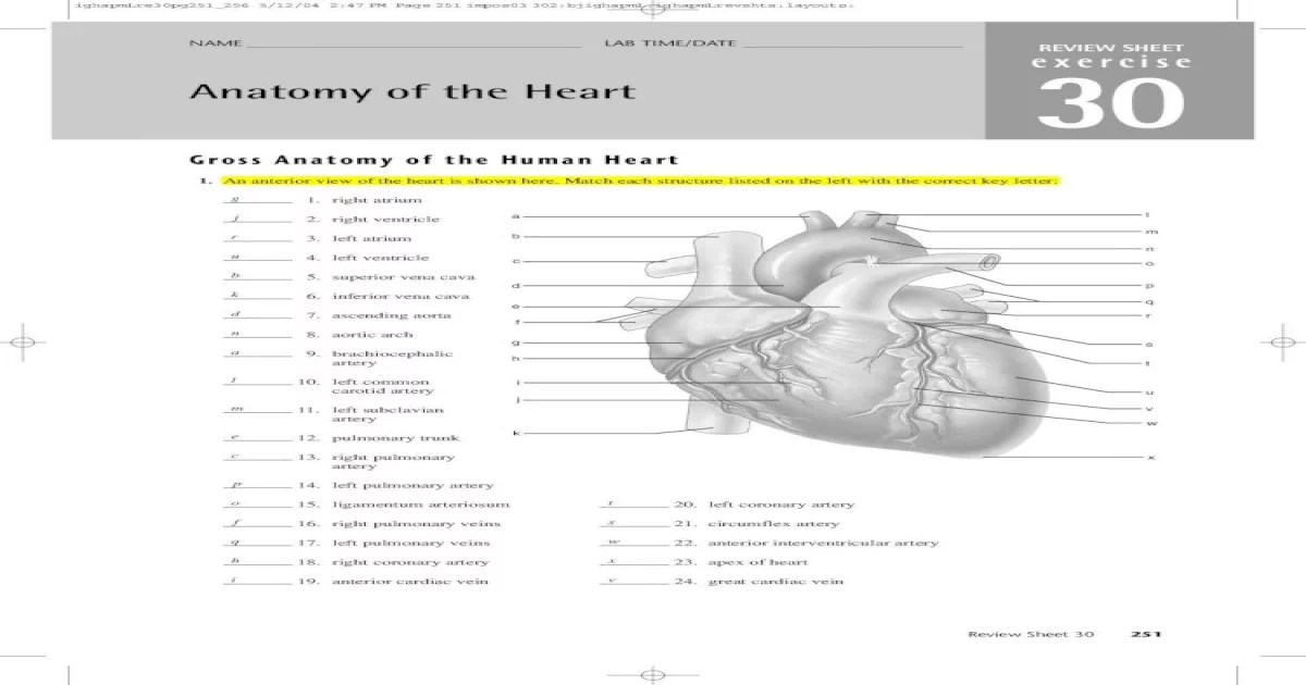
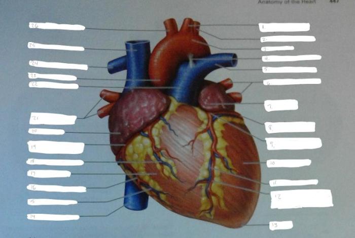
Major blood vessels connected to the heart include the superior and inferior vena cava, which bring deoxygenated blood to the right atrium; the pulmonary artery, which carries deoxygenated blood to the lungs; the pulmonary veins, which bring oxygenated blood back to the left atrium; and the aorta, which carries oxygenated blood away from the left ventricle to the rest of the body.
External Anatomy of the Heart
The external surface of the heart exhibits several distinct features. The apex, or the pointed tip of the heart, is located at the bottom of the left ventricle. The base, or the broader end of the heart, is located at the top and includes the atria and the origins of the major blood vessels.
The pericardium, a double-layered sac, surrounds the heart. The outer layer, the fibrous pericardium, provides structural support and protection. The inner layer, the serous pericardium, secretes a fluid that lubricates the heart’s surface, reducing friction during contraction.
The coronary arteries, which arise from the aorta, supply oxygenated blood to the heart muscle itself.
Internal Anatomy of the Heart
The interior of the heart consists of the atrioventricular valves (tricuspid valve on the right and mitral valve on the left) and the semilunar valves (pulmonary valve on the right and aortic valve on the left). These valves prevent backflow of blood and ensure unidirectional flow through the heart.
The electrical conduction system of the heart, which initiates and coordinates heart contractions, consists of the sinoatrial node (SA node), the atrioventricular node (AV node), and the bundle of His. The SA node, located in the right atrium, generates electrical impulses that spread through the heart, causing the atria to contract.
The AV node delays the impulses slightly, allowing the atria to fill completely before the ventricles contract.
The coronary sinus, a large vein, collects deoxygenated blood from the heart muscle and drains it into the right atrium.
Blood Flow through the Heart
Blood flow through the heart follows a specific pathway. Deoxygenated blood from the body enters the right atrium via the superior and inferior vena cava. It then passes through the tricuspid valve into the right ventricle, which pumps the blood to the lungs via the pulmonary artery.
Oxygenated blood returns to the heart via the pulmonary veins and enters the left atrium. From there, it passes through the mitral valve into the left ventricle, which pumps the blood to the rest of the body via the aorta.
The cardiac cycle refers to the sequence of events that occur during one heartbeat. It consists of systole (contraction) and diastole (relaxation) phases for both the atria and ventricles.
Heart rate and blood pressure are regulated by various mechanisms, including the autonomic nervous system, hormones, and intrinsic cardiac properties.
Clinical Significance, Review sheet 30 anatomy of the heart
Understanding heart anatomy is crucial for diagnosing and treating cardiovascular diseases. Abnormalities in heart structure or function can lead to various conditions, including congenital heart defects, coronary artery disease, heart failure, and arrhythmias.
Knowledge of heart anatomy enables healthcare professionals to accurately interpret diagnostic tests such as echocardiograms and electrocardiograms, which provide valuable information about the heart’s structure and function.
Furthermore, understanding heart anatomy is essential for performing surgical interventions such as heart valve replacement, coronary artery bypass grafting, and heart transplantation.
FAQ Explained
What are the four chambers of the heart?
The four chambers of the heart are the right atrium, right ventricle, left atrium, and left ventricle.
What is the function of the coronary arteries?
The coronary arteries supply oxygen-rich blood to the heart muscle.
What is the electrical conduction system of the heart?
The electrical conduction system of the heart is responsible for generating and transmitting electrical impulses that coordinate the heart’s contractions.


