
The microscope images indicate that ovalbumin possesses unique structural characteristics and morphology, providing valuable insights into its molecular composition and biological functions.
Microscopy techniques offer a powerful tool to visualize and analyze the intricate details of ovalbumin, enabling researchers to gain a deeper understanding of its properties and interactions.
Overview of Ovalbumin
Ovalbumin is a major protein found in egg white, constituting approximately 54% of its total protein content. It belongs to the family of albumin proteins, known for their solubility in water and ability to coagulate upon heating. Ovalbumin is a globular protein with a molecular weight of approximately 45 kDa.
It is composed of a single polypeptide chain consisting of 385 amino acids. Ovalbumin is characterized by its high nutritional value, containing all the essential amino acids required for human health.
Microscopic Examination of Ovalbumin
Microscopy techniques provide valuable insights into the structure and morphology of ovalbumin. Bright-field microscopy and transmission electron microscopy (TEM) are commonly employed to visualize ovalbumin under a microscope.
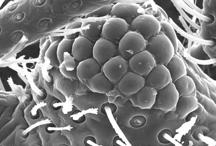
Bright-Field Microscopy, The microscope images indicate that ovalbumin
Bright-field microscopy is a simple and widely used technique for observing ovalbumin. It involves illuminating the sample with white light and using a compound microscope to magnify the image. Under bright-field microscopy, ovalbumin appears as bright, spherical or oval-shaped structures against a dark background.
The size and shape of ovalbumin can be estimated using this technique.
Transmission Electron Microscopy (TEM)
TEM is a more powerful microscopy technique that provides detailed structural information about ovalbumin. In TEM, a beam of electrons is transmitted through the sample, and the resulting image is formed by the interaction of electrons with the sample’s atoms.
TEM allows for higher magnification and resolution compared to bright-field microscopy, revealing the ultrastructure of ovalbumin, including its internal components and surface features.
Ovalbumin’s Appearance under the Microscope

Under bright-field microscopy, ovalbumin appears as spherical or oval-shaped structures with a smooth surface. The size of ovalbumin can vary depending on the preparation and conditions, typically ranging from 50 to 100 nm in diameter. TEM images of ovalbumin reveal a more detailed structure, showing a dense core surrounded by a less dense shell.
The core of ovalbumin contains a high concentration of protein molecules, while the shell consists of carbohydrates and other molecules.
Interpretation of Microscope Images
The microscopic features of ovalbumin provide insights into its molecular structure, interactions, and biological functions. The size and shape of ovalbumin can indicate its aggregation state and interactions with other molecules. The presence of internal components or surface features can suggest the presence of specific binding sites or conformational changes.
Advanced Microscopy Techniques for Ovalbumin Analysis

In addition to bright-field microscopy and TEM, advanced microscopy techniques can provide further insights into the structure and dynamics of ovalbumin. Atomic force microscopy (AFM) is a non-invasive technique that allows for the visualization of the surface topography of ovalbumin at the nanoscale.
Fluorescence microscopy, combined with specific fluorescent probes, can provide information about the localization and interactions of ovalbumin within cells or tissues.
Case Studies or Examples
Microscopy has been extensively used to analyze ovalbumin in various research and industrial applications. In one study, TEM was employed to investigate the structural changes of ovalbumin upon heating, providing insights into its denaturation process. In another study, AFM was used to characterize the surface properties of ovalbumin nanoparticles, revealing their potential for drug delivery applications.
Helpful Answers: The Microscope Images Indicate That Ovalbumin
What are the key features of ovalbumin under the microscope?
Ovalbumin appears as oval or spherical structures under the microscope, exhibiting a smooth surface and a well-defined shape.
How can microscopy help determine the molecular structure of ovalbumin?
Microscopy techniques, such as electron microscopy, provide high-resolution images that allow researchers to visualize the arrangement of atoms and molecules within ovalbumin, revealing its molecular structure.
What are some applications of microscopy in ovalbumin research?
Microscopy is used in various applications, including studying the interactions of ovalbumin with other proteins, analyzing its conformational changes, and investigating its role in biological processes.
